Nerve Block/Pain Relief Manikin
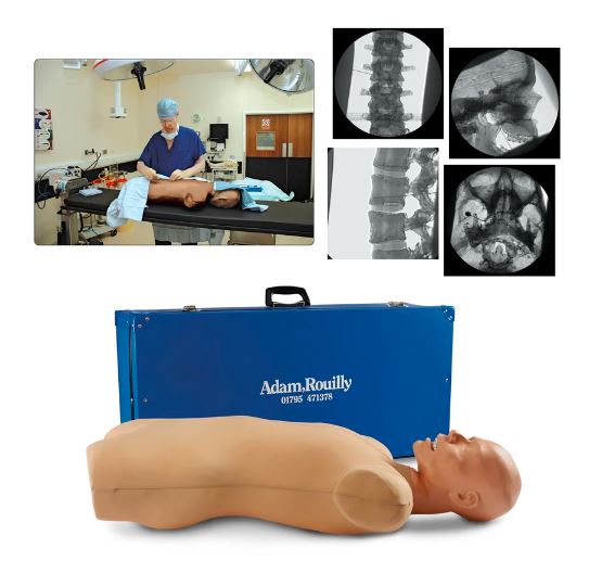
Teaches correct needle placement in nerve blocks for pain management under X-ray image intensifier control.
Students will be able to use their knowledge of surface anatomy to identify the appropriate locations for various nerve blocks, the point of needle insertion, the angle of direction, and how to withdraw the needle and direct it to alter the angle.
Students will also learn how to recognize contact with deep bony structures and how to employ radiological screening to ensure proper needle placement, including image orientation, identification of appropriate radiographic landmarks, and the end point of simulation to obtain the correct radiographic appearance.
Use the manikin to teach the following nerve blocks:
- Coeliac nerve block
- Cervical facet joint injection
- Cervical, thoracic, and lumbar facet joint injection and radio frequency denervation of posterior primary ramus
- Epidural injections at all spinal levels
- Lumbar sympathetic block
- Sacroiliac joint injection
- Splanchnic nerve block
- Superior hypogastric nerve block
- Trigeminal ganglion block or radio frequency needle placement
Manikin consists of a specially coated plastic human skeleton head covered in artificial skin and flesh-colored, fabric-covered torso.
Low X-ray density of the manikin reduces the doses of radiation used during simulated procedures.
Includes 38-1/2 in. L x 17-3/4 in. W x 13-3/4 in. H manikin, 4-minute DVD and leaflet illustrating the use of the manikin, and a rigid carrying case.
Two-year warranty.


